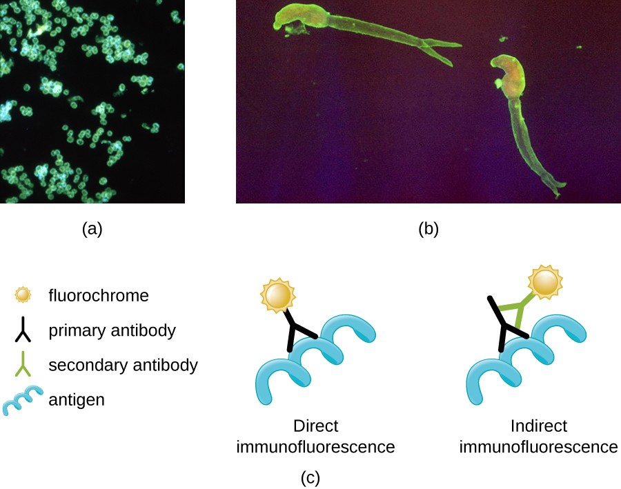
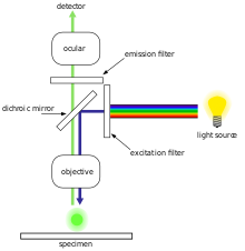
Around 1962 Ploem started work in collaboration with Schott on the development of dichroic beamspitters for reflection of blue and green light for fluorescence microscopy using epi-illumination. By combining antibodies that are labeled with different fluorochromes the location of several proteins can simultaneously be visualized in one sample.
Instruments Of Microscopy Microbiology
These flourchromes when exited with fluorescent light of given wavelength emit light with longer wavelength.
Fluorescence microscopy with multiple fluorochromes. Minimization of pixel shift in multiple fluorescence exposures using fluorescence filters from. Filter set in modern filter modules Contents with light trap 1. Current filter sets from Carl Zeiss 2.
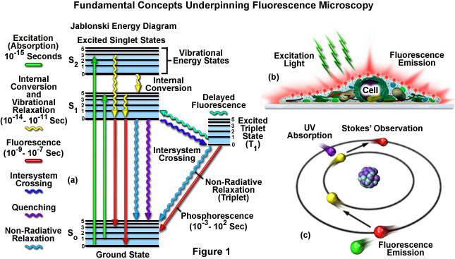
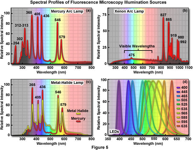
Fluorescence is a member of the ubiquitous luminescence family of processes in which susceptible molecules emit light from electronically excited states created by either a physical for example absorption of light mechanical friction or chemical mechanism. UV excitation as used traditionally for fluorescence microscopy was not optimally suited for detecting multiple fluorochromes simultaneously in a cell. Standard fluorochromes and recommended filter sets 3.
BV421 and BV480 fill a gap in the visible spectrum. Choose A Filter Cube. BD Biosciences and Chroma Technology have collaborated to introduce these fluorochromes to help advance fluorescence microscopy.
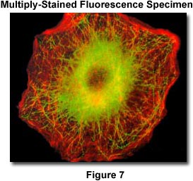
Common of these fluorochromes. Immunofluorescence microscopy is a unique method to reveal the spatial location of proteins in tissues and cells. Fluorescence With Multiple Fluorochromes This interactive tutorial explores the effects of fluorescence cube filtration of excitationemission spectra on specimens stained with multiple fluorochromes.
Proteins of interest can be marked with such fluorochromes via antibody staining or tagging with fluorescent proteins. VioDyes family Vio and Vio Bright Dyes represent a family of fluorochromes for flow cytometry and fluorescence microscopy. To operate the tutorial first use the pull-down menu to choose a sample.
They are characterized by high fluorescence intensities and low spillover making them an ideal choice for multicolor applications. Chroma Technology recognized the opportunity to make 5-color imaging easily accessible to anyone working with a basic fluorescence microscopy stand. Despite the numerous advances made in fluorescent dye synthesis during the past few decades there is very little solid evidence about molecular design rules for developing new fluorochromes particularly with regard to matching absorption spectra to available confocal laser excitation wavelengths.
When multiple fluorochromes are used there is often a fluorescence interference due to the overlapping emission spectra which is most prominent in fluorochromes that emit at adjacent wavelengths. In a sample through the use of multiple staining different probes can simultaneously identify several target molecules. Next use your mouse cursor to select a fluorescence filter cube using the radio buttons.
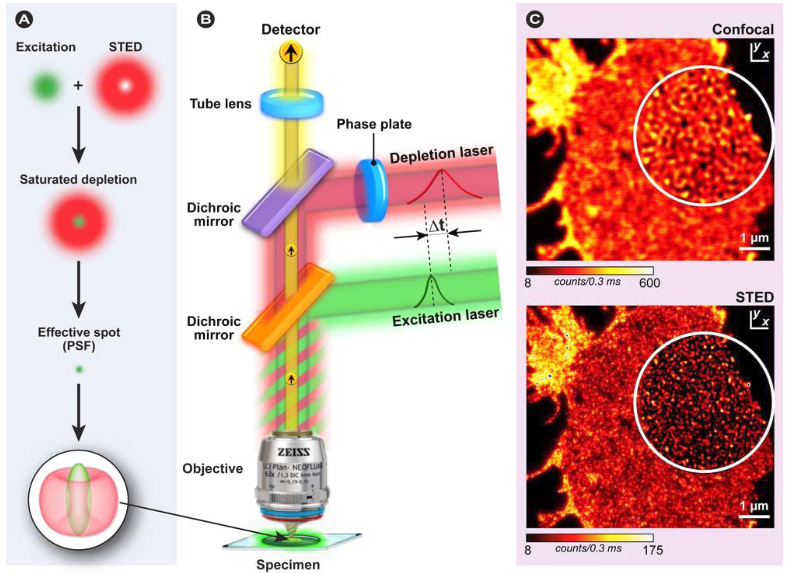
Fluorescence microscope can reveal the presence of a single fluorescing molecule. Generation of luminescence through excitation of a molecule by ultraviolet or visible light photons is a phenomenon termed. Fluorescence imaging in its conventional form reveals biological structures and processes but is limited by the number of fluorochromes that can be distinguished in one measurement.
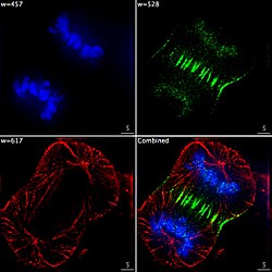
Although the fluores-cence microscope cannot provide spatial resolution below the diffraction limit of the respective objects the de-. In the case of dual- or multiple fluorchromes the data on the different wavelengths is collected separately and assigned different colors. Fluorescence microscopy reveals the distribution of fluorochromes in biological specimens whether they are introduced externally as probes or occur naturally as chlorophylls and porphyrins.
For example fluorescein isothiocyanide FITC and phycoerythrin PE emit at 520 and 576 nm respectively. This interactive tutorial explores the effects of fluorescence cube filtration of excitationemission spectra on specimens stained with multiple fluorochromes. The bleed-through effect is extremely relevant when imaging multiple fluorochromes or fluorescence proteins since the signals from one specific structure or protein will be captured in the adjacent filter set and will mask signals from both proteins or structures.
A computer generates the image from this fluorescence data. Fluorescence Microscopy Fluorescence microscope uses fluorescent light to excite fluorescent specimens. More recently the development of nanometer-sized fluorescent semiconductor quantum dots has provided a new avenue for research in confocal and widefield fluorescence microscopy.
Images of the separate fluorochromes or a merge image.
Fluorescence Microscope Wikipedia
Zeiss Microscopy Online Campus Microscopy Basics Fluorescence Microscopy

Multicolor Fish With Three To Five Fluorochromes Using Confocal Download Scientific Diagram
Molecules Free Full Text Advanced Fluorescence Microscopy Techniques Frap Flip Flap Fret And Flim Html
![]()
Confocal Microscopy Fluorophores For Confocal Microscopy Olympus Ls

Multiple Fluorochrome Analysis Of Sk N Sh Cells Employing Digital Download Scientific Diagram
Fluorescence Microscopy Fluorescence Filters Olympus Ls
Zeiss Microscopy Online Campus Microscopy Basics Fluorescence Microscopy

Multicolor Fish With More Than Five Fluorochromes Using Confocal Download Scientific Diagram

Multi Wavelength Epi Illumination In Fluorescence Microscopy Learn Share Leica Microsystems

Why Understanding Fluorochromes Is Important In Flow Cytometry Cheeky Scientist
Fluorescence Microscope Wikipedia
Ppt 7 1 Fluorochromes Fluorescence Microscopy Differentiates Between Two Kinds Of Fluorochromes Powerpoint Presentation Id 3722168

Why Understanding Fluorochromes Is Important In Flow Cytometry Cheeky Scientist